CBCT 3D
X-Rays
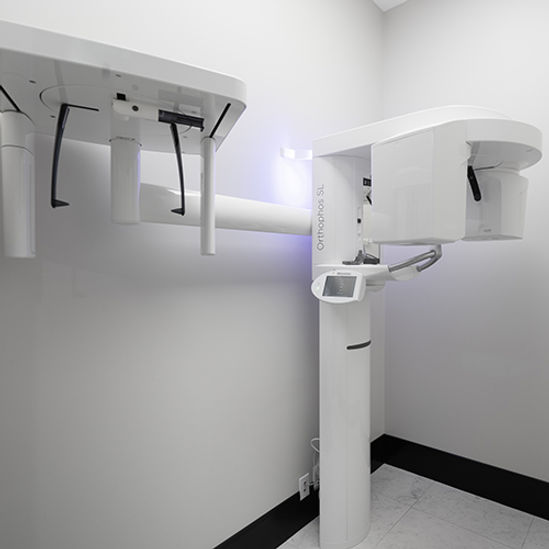
With the computer age upon us, new technology is available to allow monitoring, diagnosis, and treatment of dental problems to become faster, easier, and more thorough. Today, 3-D cone beam computed tomography (CBCT) is one such advancement that has become available to clinical dental practitioners in the last decade.
The use of 3-D cone beam technology allows dental professionals to image the head or skull area to the third dimension. Cone-beam computed tomography systems (CBCT) are a variation of traditional computed tomography (CT) systems. The CBCT system rotates around the patient, capturing data using a cone-shaped X-ray beam. This data is used to reconstruct a three-dimensional (3D) image of the following regions of the patient’s anatomy: dental (teeth); oral and maxillofacial region (mouth, jaw, and neck); and ears, nose, and throat (“ENT”).
Dental Applications
The amount of radiation exposure from a full mouth series of x-rays is equal to the amount a person receives in a single day from natural sources. We take necessary precautions to limit your exposure to radiation when taking dental x-rays. These precautions include using a lead apron shield with a thyroid collar for your protection.